
Atypical bead-cultured pearls are, by definition, cultured with unconventional beads as opposed to the typical round, predominantly freshwater shell beads. Examples of such atypical bead nuclei are irregularly shaped pieces of shell, ceramic, plastic, wax, coral beads, or even cultured or natural pearls.
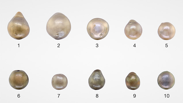
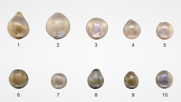
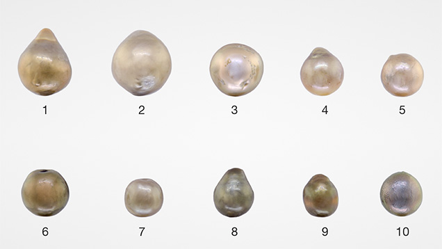
Recently, GIA’s New York laboratory received 10 loose undrilled pearls for identification. These samples exhibited a brownish bodycolor and measured from 7.17 × 6.92 × 6.42 mm to 12.25 × 9.72 × 9.28 mm (figure 1). Many were unusually lightweight for their size. Microradiographic examination of seven of the pearls revealed atypical bead nuclei (figure 2). The X-ray transparencies of the bead nuclei allowed us to observe faint demarcations and drill holes with varying degrees of clarity in the nuclei.
These atypical bead nuclei permitted some unusual nacre growth to develop within the drill holes, as shown in figure 2C, where the nacre secretion flows into the drill hole. This interesting observation concerning nacre viscosity was also described in recent work on atypical bead culturing experiments (K. Scarratt et al., “Atypical ‘beading’ in the production of cultured pearls from Australian Pinctada maxima,” GIA Research & News, Feb. 13, 2017).
To better visualize the unusual nacre growth within the drill hole of the bead in one of the pearls, we used computed X-ray microtomography (μ-CT) and subsequently reconstructed the pearl using computer software (figure 3). The reconstructed image displayed a surface indentation aligned with the bead’s drill hole (shown in orange in figure 3, left). Additional nacre growth (shown in green, figure 3, right) filled about half of the drill hole. The yellowish outer region in figure 3 (left) represents the outer nacre of the pearl.
Although this is not the first time GIA has received pearls cultured using X-ray transparent bead nuclei (Fall 2011 Lab Notes, pp. 229–230), the unusual nacre growth pattern within some of the drill holes in this sample group is worthy of note. Three-dimensional rendering using computer software and μ-CT data sets also proved very useful in visualizing the internal structure, further demonstrating the technique’s worth as a powerful tool in pearl identification.
Atypical bead-cultured pearls are, by definition, cultured with unconventional beads as opposed to the typical round, predominantly freshwater shell beads. Examples of such atypical bead nuclei are irregularly shaped pieces of shell, ceramic, plastic, wax, coral beads, or even cultured or natural pearls.
Recently, GIA’s New York laboratory received 10 loose undrilled pearls for identification. These samples exhibited a brownish bodycolor and measured from 7.17 × 6.92 × 6.42 mm to 12.25 × 9.72 × 9.28 mm (figure 1). Many were unusually lightweight for their size. Microradiographic examination of seven of the pearls revealed atypical bead nuclei (figure 2). The X-ray transparencies of the bead nuclei allowed us to observe faint demarcations and drill holes with varying degrees of clarity in the nuclei.
These atypical bead nuclei permitted some unusual nacre growth to develop within the drill holes, as shown in figure 2C, where the nacre secretion flows into the drill hole. This interesting observation concerning nacre viscosity was also described in recent work on atypical bead culturing experiments (K. Scarratt et al., “Atypical ‘beading’ in the production of cultured pearls from Australian Pinctada maxima,” GIA Research & News, Feb. 13, 2017).
To better visualize the unusual nacre growth within the drill hole of the bead in one of the pearls, we used computed X-ray microtomography (μ-CT) and subsequently reconstructed the pearl using computer software (figure 3). The reconstructed image displayed a surface indentation aligned with the bead’s drill hole (shown in orange in figure 3, left). Additional nacre growth (shown in green, figure 3, right) filled about half of the drill hole. The yellowish outer region in figure 3 (left) represents the outer nacre of the pearl.
Although this is not the first time GIA has received pearls cultured using X-ray transparent bead nuclei (Fall 2011 Lab Notes, pp. 229–230), the unusual nacre growth pattern within some of the drill holes in this sample group is worthy of note. Three-dimensional rendering using computer software and μ-CT data sets also proved very useful in visualizing the internal structure, further demonstrating the technique’s worth as a powerful tool in pearl identification.